Kaiser Permanente research ties presence of breast arterial calcification to increased risk of cardiovascular disease
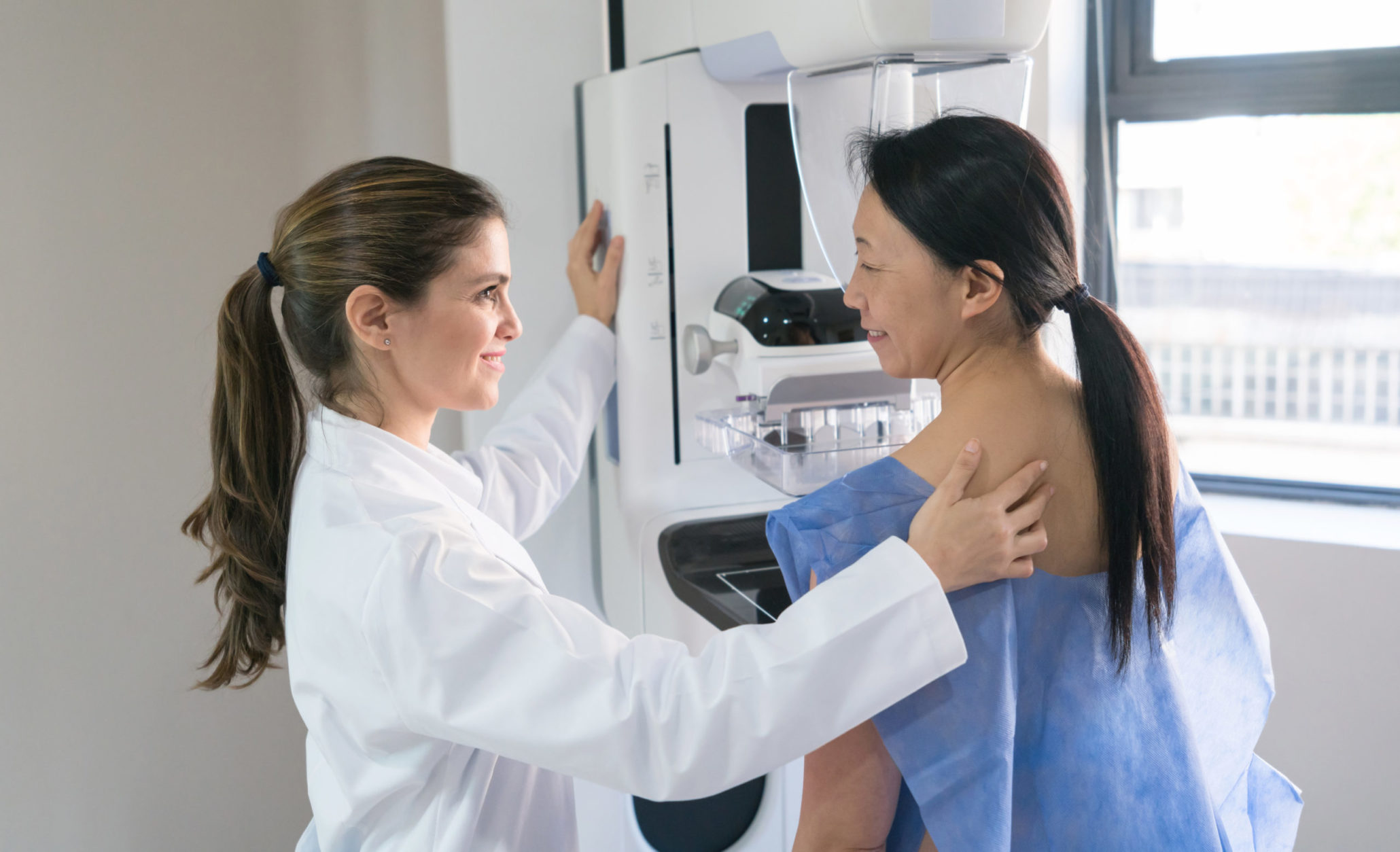
Screening mammography is used to look for early indicators of breast cancer. But a new collaborative study between Kaiser Permanente and University of California, Irvine (UCI) shows mammograms can also help determine a woman’s risk of having a heart attack or stroke or developing cardiovascular disease.

“Our findings add to the body of evidence that suggests breast arterial calcification seen on a mammogram of a postmenopausal woman provides additional information that can be used to assess her heart health,” said Carlos Iribarren, MD, MPH, PhD, a research scientist at the Kaiser Permanente Division of Research.
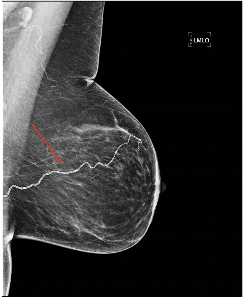
Breast arterial calcifications are calcium deposits that show up on a mammogram as white parallel lines in the breast’s arteries. These deposits, which become more common as a woman gets older, have been shown to be associated with diabetes, high blood pressure, and inflammation; they are not early indicators of breast cancer. Radiologists are currently not required to report whether these calcifications are seen on a mammogram.
The study, published March 15 in Circulation: Cardiovascular Imaging, included 5,059 female members of Kaiser Permanente Northern California between age 60 and 79 who had routine mammography screening between 2012 and 2015. After the images were reviewed for signs of breast cancer, they were sent to the research team at UCI who assessed whether and how much breast arterial calcification was present. The research team then followed the women for 6.5 years, reviewing their electronic medical records for diagnoses of a heart attack, stroke, or atherosclerotic cardiovascular disease — a collection of diseases caused by plaque buildup, such as acute coronary syndrome and peripheral arterial disease.
The study found that women who had breast arterial calcification were 51% more likely to be diagnosed with a heart attack or stroke.
Of the 5,059 women in the study, 1,338 (26.5%) were found to have breast arterial calcification. Over the 6.5-year follow-up period, 155 women were diagnosed with a heart attack or stroke and 427 with any form of cardiovascular disease. The study found the women who had breast arterial calcification were 51% more likely to be diagnosed with a heart attack or stroke, and 23% more likely to develop any form of cardiovascular disease.
The research team also used a risk calculator developed by the American Heart Association and the American College of Cardiology to assess each woman’s 10-year risk of developing heart disease. They found that 12.1% of the women with breast arterial calcifications had a high 10-year risk compared with 5.5% of the women who had no calcifications. They also identified women with low-risk scores who had breast arterial calcification, suggesting their risk could be higher than their score suggested.
“Our study has moved the needle in support of revising guidelines for reporting breast arterial calcification on mammography and incorporating this information into the currently used heart disease calculators,” said Iribarren.
This is the first prospective study looking at the association between these calcifications and heart disease to use state-of-the-art digital mammography and quantify the amount of breast arterial calcification present. It took more than 3 years for a team of radiologists to carefully review the more than 20,000 mammography images (each woman had 2 images taken of each breast) used in this study.
“We now know that arterial breast calcifications can provide additional information about a woman’s cardiovascular risk,” said the study’s senior author Sabee Molloi, PhD, a professor of radiological sciences at the UCI School of Medicine. “But to make this knowledge useful and easy to include on a woman’s mammography report, we will need to find a way to automate and standardize how we evaluate the amount of breast arterial calcification present.”
Looking at breast arterial calcification

Cardiovascular diseases — diseases that affect the heart and blood vessels — are the leading cause of death in women in the U.S. Breast arterial calcification occurs in the middle layer of the arterial wall. It is not the same as the calcification that occurs in the inner layer of the arteries (the layer that is in contact with the blood) that is seen in smokers and people with high cholesterol levels.“
“These arteries become rigid, like a hose filled with water,” Iribarren explained. “Since it occurs more often in older women, and is associated with diabetes and high blood pressure, it’s a sign that there are biological pathways in the body that impact the propensity to develop calcification and that increase risk for heart disease. Our research team plans to explore this biological process to identify what may cause this to occur in the breast arteries.”
The new research is part of the MINERVA study (MultIethNic Study of BrEast ARterial Calcium Gradation and CardioVAscular Disease) Iribarren and colleagues launched in 2010. Previous MINERVA analyses showed breast arterial calcification was associated with the ankle brachial index (a measure of peripheral arterial disease) but not with mild cognitive impairment or dementia or bone mineral density. Going forward, Iribarren and Molloi plan to investigate whether breast arterial calcification is also helpful in predicting cardiovascular disease among younger women, ages 40 through 59.
“Because mammography is a routine part of a woman’s health care, it makes sense to find a way to use the information it provides about heart disease risk,” said Iribarren.
This study was funded by the National Heart, Lung, and Blood Institute.
Co-authors include Malini Chandra, MS, Catherine Lee, PhD, and Gabriela Sanchez, MA, of the Division of Research; Danny L. Sam, MD, of The Permanente Medical Group; Farima Faith Azamian, MD, and Huanjun Ding, PhD, and Nathan D. Wong, PhD, of the University of California, Irvine School of Medicine; and Hyo-Min Cho, PhD, of the Korea Research Institute of Standards and Science.
# # #
About the Kaiser Permanente Division of Research
The Kaiser Permanente Division of Research conducts, publishes and disseminates epidemiologic and health services research to improve the health and medical care of Kaiser Permanente members and society at large. It seeks to understand the determinants of illness and well-being, and to improve the quality and cost-effectiveness of health care. Currently, DOR’s 600-plus staff is working on more than 450 epidemiological and health services research projects. For more information, visit divisionofresearch.kaiserpermanente.org or follow us @KPDOR.


This Post Has 0 Comments